Scientists dig up proteins from the past

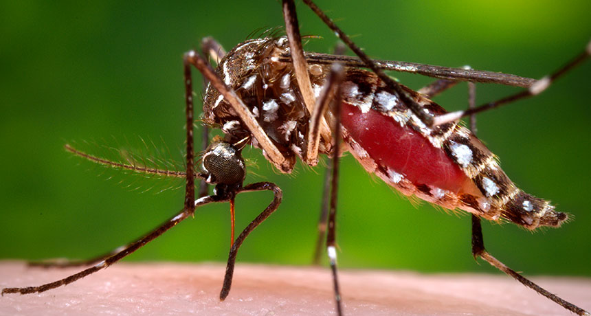
The influenza virus is a quick-change artist. In a few decades, its genome can evolve as much as animal genomes can over millions of years. That means that the viral proteins, including those that alert our bodies to an infection, constantly reinvent themselves, threatening our immune systems and frustrating vaccine developers.

For Jesse Bloom, a biologist studying how evolution affects proteins, that relentless change is an opportunity. Thanks to data collected during past flu seasons, Bloom knows the exact genetic makeup of some ancestors of today’s influenza viruses. His lab group at the Fred Hutchinson Cancer Research Center in Seattle uses that information to figure out how the viruses made their immunity-dodging transformations.

Bloom and others are part of a growing group of scientists who practice “evolutionary biochemistry.” They seek to explain life’s tremendous diversity and determine exactly how that diversity emerged. Rather than focusing on how plants or animals adapted to different environments, however, these researchers consider diversity on a much smaller scale: Their work aims to explain how the small set of proteins that powered primitive life-forms evolved into the millions of specialized proteins that drive biological processes today.

Exploiting the genetic records, Bloom can assemble virus proteins that existed in bygone times, then reconstruct how they evolved, one amino acid at a time. Other researchers are analyzing modern species to resurrect the ancestral forms of biological molecules that have evolved over millions of years.

With a historical protein in hand, researchers can test how swapping out a single amino acid — as evolution might have done — changes how the protein flexes or folds and connects (or doesn’t) with other molecules. By trying out alternate versions of a protein’s history through stepwise amino acid changes, scientists can learn how a protein’s physical form has both enabled and constrained its evolution.

Ultimately, this work might answer some long-standing questions: To what extent does evolution depend on chance events? Can evolution reach the same point by traveling different paths? How does biological complexity evolve? Such experiments are also helping researchers who study modern proteins sort out how the order of amino acids relates to biological function.
That ordered series of amino acids is spelled out by the gene that holds the blueprint for a protein. Once the proper amino acids are strung together, they origami-fold into tiny structures with nooks and protrusions that determine what the protein does inside a cell. A protein’s folded shape lets it grab on to specific bits of DNA or hasten certain chemical reactions. Mutations in a gene can shift the resulting protein’s shape or alter subtle aspects of its behavior so that, over time, a protein’s function can change. But the possibilities are not endless. New proteins that fall apart, fail to fold or don’t perform as needed don’t survive the tests of natural selection.

“The physical determinants of folding, stability, solubility, function and specificity are absolutely essential aspects of the evolutionary process,” says University of Chicago biologist Joe Thornton. “That has not been widely appreciated or explicitly addressed until pretty recently.” Now, Thornton says, it’s clear that to understand molecular evolution, it’s important to study proteins as functioning, physical objects.

As they reconstruct proteins’ pasts, researchers are finding that genetic mutations sometimes remodel a molecule just enough to give a chance to other mutations that would have failed earlier. That creates opportunities for new features and functions to evolve — an idea that biologists have considered for decades but have only just begun to explore in the lab.

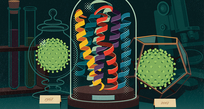
Bloom and colleagues, for instance, used an influenza virus protein called nucleoprotein to examine how interactions among mutations have affected the overall evolution of the virus. Understanding the combined effects of several mutations could allow researchers to anticipate the short-term effects of new genetic variation. That knowledge could help improve forecasts of which viral strains are likely to circulate in upcoming flu seasons, important information for designing effective vaccines.

Comparing nucleoprotein genes from strains of the virus isolated in 1968 and 2007, Bloom’s team mapped out the most likely steps by which the 1968 protein morphed into its newer form. Though nucleoprotein still plays the same role that it did in 1968 — aiding in the assembly of viral RNA — 33 of its 498 amino acids changed over those four decades, and a few changed more than once, the researchers reported in 2013 in eLife.

Bloom’s team built the 1968 nucleoprotein, then tested the effects of introducing each historical mutation. Some of the mutations affected parts of the protein that tip off a person’s immune cells that an invader is present — they probably helped the flu virus avoid detection. But on their own, some of those changes were bad for the virus: The nucleoprotein could no longer stay properly folded long enough to do its job.

During the course of the nucleoprotein’s evolution, some mutations boosted the protein’s stability, giving it a bit of a buffer. When later mutations occurred, allowing the virus to buck immune recognition, these earlier changes probably held the structure stable so the protein could still function.

When a mutation’s effects depend on other mutations, this interplay is called epistasis. These interactions within individual molecules have been important in shaping evolutionary trajectories, says University of Oregon biophysicist Michael Harms, who is studying how diverse functions evolved in a group of proteins called s100s. He calls epistasis “the common feature in all of evolution.”

Codependent interactions don’t occur just between pairs of mutations. They can be significantly more complex. Analyzing data from other labs, Harms has found epistatic interactions involving up to six different mutations. Such interplay means that in many cases, if genes had transformed themselves just a bit differently, evolution would have veered onto a different course.

Green light
Scientists call mutations that lay the groundwork for future change “permissive” mutations. Some protein functions came about only after permissive mutations modified an evolving molecule in highly improbable ways.

Thornton uses ancestral protein reconstruction to study how steroid hormones — which control stress responses, growth and sexual developmental in vertebrates — evolved partnerships with their receptors. Receptors are proteins that bind to specific partners to activate responses in the cell. By comparing steroid receptors in different species, Thornton can map the evolutionary relationships between the molecules and infer the likely amino acid sequence of their common ancestor. Then he introduces a DNA molecule that encodes the long-extinct protein into lab-grown cells. Those cells use the genetic instructions to manufacture a tiny piece of the deep past.

Many of Thornton’s studies begin with a 450-million-year-old receptor protein that he and colleagues reconstructed in 2006. The protein gave rise to modern receptor molecules that are activated by different hormones. One receptor, the glucocorticoid receptor, responds to the stress hormone cortisol. The other, the mineralocorticoid receptor, controls levels of salt and other electrolytes in response to the hormone aldosterone. Thornton’s team found that their reconstructed ancestor could be activated by both cortisol and mineralocorticoids.

A receptor that responded only to cortisol appeared 40 million years after the promiscuous receptor, Thornton showed. His team found a set of amino acid changes that converted the general ancestral receptor into the cortisol-specific one. But the mutations that changed the ancient receptor’s preference couldn’t have generated a functional receptor by themselves, experiments showed.

“The function-switching mutations are not tolerated on their own,” Thornton says. They destabilize parts of the receptor. Like the flu virus’s evolving nucleoprotein, the ancestral receptor’s structure had to be buttressed before it could withstand the mutations that would make the receptor choosier.

Two amino acid changes quietly readied the ancient receptor for its transformation, Thornton and colleagues reported in 2009 in Nature. Without them, the path to the function-switching mutation would have been inaccessible. “If we were to wind back the clock and set history rolling again, it’s very unlikely that those permissive mutations would occur,” he says. “We would have ended up with a very different glucocorticoid receptor and a very different endocrine system.”

Thornton and Harms, then a postdoctoral researcher in Thornton’s lab at University of Oregon in Eugene, explored whether evolution could have taken an alternate route to the same end. Harms created and screened thousands of variants of the ancestral protein, searching for alternative mutations that might have set it up for the same functional switch. He found none, the researchers reported in Nature in 2014. Evolution, it seems, had acted on a rare opportunity.

Biophysical analyses of variant receptor proteins showed why so few mutations enabled cortisol-specific binding to evolve. Although certain parts need extra support, the receptor also needs to be able to transition between two forms: an inactive conformation when no cortisol is present, and a gene-activating conformation when the hormone binds. Some mutations stabilize the active form of the receptor too much, locking it into an “always-on” configuration. Mutations also had to be compatible with the ancestral protein on their own, before the function-switching mutations were introduced.

“A mutation has to fulfill all these requirements, and that is not easy to do,” Thornton says. “That seems to be the explanation for why permissive mutations [for this functional switch] are so rare.”

But not every new function is the result of complicated epistatic interactions. In January in eLife, Thornton and Ken Prehoda of the University of Oregon described an ancient protein that gained a completely new function by way of a single amino acid change.

The team studied the origins of an animal protein that helps cells orient themselves in space before dividing. Doing so is vital for positioning new cells in the right places within a growing body. Single-celled life-forms had to get this right before multicellular organisms could evolve.
Thornton, Prehoda and colleagues focused on a segment of the protein called GK PID (for GK protein-interaction domain), which orients cells by acting as a scaffold during division. The billion-year-old ancestor of GK PID did nothing of the sort. It was an enzyme predecessor to the modern guanylate kinase, which catalyzes a chemical reaction that cells use to make some of the building blocks of DNA. Amazingly, Thornton says, one mutation was enough to transform the ancestral protein from an enzyme to a working scaffold.
That surprising result is an example of why developing general theories about the physical principles shaping evolution requires a grasp of the evolutionary histories of a broader collection of proteins.

“Every time people take [a protein] apart, they see a new feature,” Harms says. Fortunately, he says, thanks to faster computers, better software and a growing number of genomes to reference, research on ancestral protein reconstruction is on the rise.

Roads taken
While chance events can shift the landscape of evolution’s possibilities, evolving proteins also have some freedom to explore. They can take more than one path to some functions.

Douglas Theobald, a biochemist at Brandeis University in Waltham, Mass., has seen this in his own investigations of an enzyme that many cells use to produce energy without oxygen. The enzyme, lactate dehydrogenase, evolved from structurally similar enzymes not just once, but at least four times in different groups of organisms. By reconstructing the evolutionary events that transformed a similar enzyme, malate dehydrogenase, into lactate dehydrogenase, Theobald and colleagues found that two groups of single-celled parasites came by the same enzyme in different ways. The researchers reported the findings in eLife in 2014 and in Protein Science in February.

The work demonstrates that different genetic backgrounds may steer evolution along different paths in different organisms but still lead to similar outcomes, Theobald says. “Even if there is a lot of epistasis, there’s still lots of different ways to the same function.”

Biochemist Susan Marqusee of the University of California, Berkeley has also found that there’s more than one way for a protein to do something new.

Marqusee collaborated with Thornton’s team to compare how two bacteria, Escherichia coli and the heat-loving Thermus thermophilus, evolved enzymes that do the same job at very different temperatures. T. thermophilus thrives in hot springs, at temperatures that would cause most proteins to fall apart. Biochemists are eager to borrow from nature’s strategies to engineer heat-tolerant proteins but have struggled to find general principles that account for this property. By reconstructing the common ancestor of the RNA-snipping enzyme known as H1 from E. coli and T. thermophilus, Marqusee’s team found out how the bacterial protein takes the heat.

That 3-billion-year-old common ancestor was less stable than the enzyme that T. thermophilus uses today, the team reported in 2014 in PLOS Biology. As the heat-tolerant protein evolved, its stability steadily increased — not because of any one innovation, but by virtue of distinct biophysical strategies at different points in time.

“The physical chemistry doesn’t really matter as long as in the end, they add up to the right phenotype,” Marqusee says. Because evolution was able to take advantage of different amino acids to boost stability in a variety of ways, the enzyme’s growing resilience to hot environments didn’t depend on the chance occurrence of a particular set of mutations.

Foggy future
Studies of how proteins have evolved in the past are unlikely to spell out how evolution will proceed in the future. “The emerging picture is that the role of chance is so great that long-term predictions of the future evolution of any protein is a very risky enterprise,” Thornton says. But recent research does offer insights into how and why today’s proteins do what they do.
One example comes from Thornton’s work on how the DNA-binding sites on steroid receptors have evolved along with their DNA targets. The hormone-activated receptors act as transcription factors, binding to specific sections of DNA to switch on certain genes. In 2014, Thornton’s team reported in Cell that a bulky amino acid in an ancestral protein prevented the protein from binding to the stretch of DNA favored by many of today’s steroid receptors. The ancestral protein awkwardly bumped up against the DNA, unable to make enough contact to really grab on. The receptor gained its new specificity when mutations ended those obstacles and introduced new clashes that blocked its access to the former binding site.

Researchers often can’t tell which differences between two related proteins make them behave differently. But reconstructing evolutionary paths can point them in the right direction.

Using ancestral reconstruction, Theobald and Brandeis colleague Dorothee Kern studied how Abl, a growth-promoting protein linked to chronic myelogenous leukemia, diverged from the related Src protein. The researchers wanted to know why the anticancer drug Gleevec binds to and shuts off Abl without obstructing Src, even though Src has a very similar structure. Theobald, Kern and colleagues identified 15 amino acids in Abl that are crucial for Gleevec binding. The amino acids influence how the protein transitions between two different configurations (that shape-shifting is disrupted in some patients with Gleevec-resistant cancers). The finding, published last year in Science, suggests that researchers may be able to develop better drugs by considering these conformational shifts.

Some proteins, or parts of proteins, might even be inherently more able to evolve than others. Certain parts of the fast-evolving viral protein hemagglutinin are unusually tolerant of change, Bloom and Bargavi Thyagarajan, who was a postdoctoral researcher in Bloom’s lab, reported in 2014 in eLife. Antibodies against hemagglutinin are the immune system’s best defense against influenza, but the protein is adept at escaping detection.

The researchers used a relatively new method called deep mutational scanning to build and test hemagglutinin proteins with nearly every possible amino acid change — about 10,000 in all — in viruses grown in the lab. In a host, changes that disguise hemagglutinin from the immune system would be advantageous. Even though there was no immune system to hide from in the lab, viruses still survived more changes to parts of hemagglutinin that would be recognized by an immune system than they did changes to other parts of the protein. Bloom and his graduate student Michael Doud reported a more detailed view of the protein and the areas that are more and less likely to tolerate mutations online on bioRxiv.org in April.
That’s good for the virus, but bad for people. Hemagglutinin seems capable of accumulating change in the very sites that vaccine developers would like to remain the same. But the finding also suggests that flu vaccines designed to target less mutation-tolerant regions of hemagglutinin might be more likely to protect against the flu from season to season. That’s a strategy some labs are already exploring — targeting the less-evolvable stalk of hemagglutinin’s lollipop-shaped structure.

It’s not yet clear why certain parts of the hemagglutinin protein tolerate change so well; Bloom hopes that studying the mutational tolerance of other proteins will help researchers figure that out.

“We’re never going to be able to predict evolution precisely, because it’s a highly stochastic process,” Bloom says. “But I think we can make better forecasts about many of the evolutionary processes that affect us. These are really challenging problems, but I think we are getting to the point where we can use experiments and molecular understanding to help us think about these processes.”

Winning helium hunt lifts hopes element not running out

The world’s known helium reserves just ballooned. Applying gas-finding techniques from the oil industry, scientists uncovered a vast reservoir of more than a trillion liters of helium gas beneath Tanzania. That’s enough to satisfy the world’s helium needs for around seven years, the researchers announced June 28 at the Goldschmidt Conference, a geochemistry meeting being held in in Yokohama, Japan. The find may allay fears that a global helium shortage will hit when the U.S. Federal Helium Reserve — currently the world’s largest helium source — runs dry within the next few years.

While previously known helium reserves were discovered by chance during oil and gas exploration, geologist Diveena Danabalan of Durham University in England and colleagues applied geologic know-how to their helium hunt. Helium accumulates underground during the radioactive decay of unstable elements such as uranium. That helium, though initially trapped, can be liberated when surrounding rock melts during volcanic activity. Using this information as well as seismic imaging of gas-trapping underground formations, the researchers discovered five spots in a volcanic region of Tanzania where water and helium-rich gas bubble to the surface from underground reservoirs.

The researchers predicted that they will be able to find more helium reservoirs and help meet society’s helium needs. Those needs go beyond just making balloons float and voices sound squeaky: Helium is essential for scientific research and a critical component of the cooling systems that allow medical MRI scanners to function.

Juno spacecraft is on its final approach to Jupiter

All systems are go for the Juno spacecraft’s July 4 encounter with Jupiter.

“We couldn’t be more excited about being this close to Jupiter’s doorstep,” said Diane Brown, Juno program executive at NASA Headquarters in Washington, D.C., during a June 30 news briefing.

The scientific instruments have been shut off and the final command sequence for going into orbit around Jupiter has been uploaded to the spacecraft’s computers. On July 4, the probe will fire its main engine for 35 minutes, using it as a brake to slow down and be captured by Jupiter’s gravity. Once in orbit, Juno will spend 20 months figuring out what’s hiding beneath the thick clouds that encase the planet.

Juno has been busy during its final approach. On June 28, it got one more look at Jupiter and three of its moons. And last week Juno monitored changes in interplanetary plasma (see below) as it crossed a magnetic boundary that shields Jupiter from the stream of charged particles blowing from the sun.

Now all scientists can do is wait. “I have mixed emotions,” said mission lead Scott Bolton, a planetary scientist at the Southwest Research Institute in San Antonio. “I’m excited, but I also have tension and nervousness.” Juno has to perform a critical engine burn all on its own while passing through treacherous belts of radiation that encircle the planet. A series of radio tones from the spacecraft will let mission scientists know whether or not it worked.

“Come see us on July 4,” Bolton said.

Documentary looks for meaning in Koko the gorilla’s life

For the last four decades, Koko, the world’s most famous gorilla, has lived in a trailer in Silicon Valley, the subject of the longest-running project on ape sign language. With a reported vocabulary of hundreds of signs, Koko has appeared to express feelings almost anyone can relate to — a love of kittens, a desire to be a mother.

A new PBS documentary argues that Koko’s remarkable life “challenges what it is that makes humans unique.” The problem, though, is that the film never really makes clear what “it” is. Rather than diving into the question of ape language and dissecting Koko’s abilities, Koko — The Gorilla Who Talks focuses more on the relationship between Koko and researcher Penny Patterson.
Patterson began working with Koko in 1972 while a Ph.D. student at Stanford University, with the aim of conducting the first sign language experiment with a gorilla. Koko was an infant, living at the San Francisco Zoo. By 1977, Patterson had negotiated to take ownership of Koko.

After completing her Ph.D., Patterson drifted away from mainstream science, and her relationship with Koko seems to have morphed from researcher and study subject to mother and child. Patterson appears deeply attached to Koko, and she seems to genuinely believe Koko is communicating her thoughts and feelings.
Skeptics interpret Koko’s behavior differently. Columbia University psychologist Herbert Terrace, who appears in the film, has conducted his own research on primate communication and intelligence. He suggests Koko is largely mimicking Patterson to receive rewards. Patterson, he argues, has failed to produce any data that prove otherwise.
The reality is probably somewhere in between these extremes. It’s difficult for anyone to really know what’s going on inside an animal’s head, but the idea of conversing with animals is deeply appealing. In the end, the film may reveal more about human behavior  — our infinite capacity for empathy (SN Online: 6/29/16) and our yearning to bond with others — than it does about the capabilities of Koko or any of our other ape cousins.

Risk of travelers to Olympics sparking new Zika outbreaks low

Any traveler to the Olympics could potentially bring Zika home, but just four countries bear a substantial risk of seeing the virus spread.

Chad, Djibouti, Eritrea and Yemen all have the right ingredients to sustain mosquito-borne transmission, researchers report July 13 in a weekly report of the U.S. Centers for Disease Control and Prevention. Few people typically travel from those countries to places where Zika virus is active, but the Olympics will change that.

To gauge the risk of a single person traveling to Rio de Janeiro for the Olympics, becoming infected and then sparking an outbreak back home, CDC scientist Ardath Grills and colleagues analyzed environmental and population data for 206 countries planning to participate in the Games.

All countries risk importing Zika from Rio, the authors write, but only 19 not currently reporting Zika outbreaks have the susceptible populations and environmental conditions needed to keep local transmission going. And all but Chad, Djibouti, Eritrea and Yemen already have lots of travelers trekking back and forth from Zika-afflicted countries.

For most countries, including the United States, travel to the Olympics doesn’t add much to the risk (SN Online: 6/14/16). (Overall, the number of people planning to travel to the Games — up to an estimated 500,000 — is less than 0.25 percent of the total number of travelers in 2015 to countries with Zika, the researchers estimate.)

The new analysis is based on “worst-case scenarios,” the authors say. It does not change current public health warnings: Pregnant women should steer clear of the Games and people should take steps to avoid spreading the virus via sexual transmission when they return home.

New brain map most detailed yet

Analyzing a bevy of diverse data, scientists have drawn a new map of the human brain in extreme relief. Their approach demarcated 180 areas in each half of the outer layer of the brain — including 97 regions in each half that haven’t been described before. The high-resolution map will allow scientists to more precisely scrutinize brain regions and see how they change with, for instance, age and disease.

Many previous maps of the brain have been built with just one type of data. The new map, described July 20 in Nature, forms a holistic view of the brain by combining several different types of information. These specs included how areas behaved while doing certain tasks or nothing at all, as well as detailed anatomical data about the shape and thickness of the brain. Using these metrics from 210 healthy people, neuroscientist David Van Essen of Washington University in St. Louis and colleagues found that each hemisphere contains 180 distinct areas (separated by black lines in image). In this view, colors show how tightly linked each area is to other brain areas that handle auditory (red), touch and movement (green) or visual (blue) information.

Yeasts hide in many lichen partnerships

The discovery of unknown yeasts hiding in lichens from six continents could shake up a basic idea of what makes up a lichen partnership.

For more than a century, biologists have described a lichen as a fungus growing intimately with some microbes (algae and/or cyanobacteria) that harvest solar energy. The fungus is treated as so important that its name serves as the name for the whole lichen.

Biologists have recognized that more than one fungus can show up in lichen close-ups, but their role hasn’t been clear. Now that may be on the brink of changing.
Fifty-two genera of lichens collected from around the world include a second fungus — single cells, called yeasts, of a previously unknown order now christened Cyphobasidiales. Toby Spribille of the University of Graz in Austria and colleagues report the finding online July 21 in Science.
The first example discovered illustrates why these yeasts might turn out to be more than parasites or mere hitchhikers, says study coauthor John McCutcheon of the University of Montana in Missoula. He and Spribille started the research out of curiosity. They wondered how the yellow, toxin-bearing, thready tangles of lichen called Bryoria tortuosa could have the same fungus and the same algal partner — and thus technically be the same species — as the brown, toxin-poor lichen traditionally called B. fremontii. The researchers looked to see which genes were active in each lichen in hopes that some discrepancy could explain the difference in forms. What the researchers found had nothing to do with the alga or previously known fungus. Instead, ample genetic activity of more abundant yeasts in the toxic B. tortuosa turned out to be the most striking disparity.

After five years of work, the research team now has microscope images of yeast cells embedded in the outer layer, or cortex, of B. tortuosa. Gene-activity results suggest that the yeasts could be what’s making the difference between the forms, maybe even synthesizing toxic vulpinic acid. The yeasts turning up across this widespread class of lichens might explain other mysteries, such as why researchers have largely failed to re-create lichen partnerships in the lab.
It’s a bold hypothesis, but lichenologist Robert Lücking of the Botanic Garden and Botanical Museum Berlin-Dahlem takes the idea of yeast partners seriously. “This will be a huge surprise to the lichenological and mycological community,” he says.

Distinctions blur between wolf species

Wolves are having something of an identity crisis. Gray wolves and coyotes might be the only pure wild canine species in North America, a new genetic analysis suggests. Other wolves — like red wolves and eastern wolves — appear to be blends of gray wolf and coyote ancestry instead of their own distinct lineages.

Red wolves contain about 75 percent coyote genes and 25 percent wolf genes, an international team of scientists reports online July 27 in Science Advances. Eastern wolves have about 25 to 50 percent coyote ancestry.
That finding adds another twist to the ongoing battle over wolf protection and regulation in the United States: how to protect a population that’s not its own species but carries valuable genetic information.

Gray wolves used to roam much of North America — until they were hunted to near-extinction. Protection under the Endangered Species Act has helped them to rebound, but their current range is still far smaller than it used to be. Red wolves, found in the southeastern United States, and eastern wolves, found in the Great Lakes region, look similar to gray wolves but are often treated as distinct species. The two groups occupy territory where gray wolves are now scarcer (in the Great Lakes area) or completely gone (in the southeast).

The new study examined the entire genetic makeup, or genome, of 23 wild canines from around North America. The researchers compared the mixed genomes to those from pure coyotes and Eurasian wolves to figure out what percent of each animal’s genetic material came from the wolf and what part came from the coyote.

Red and eastern wolves have historically mated with coyotes, the team found. But gray wolves have recent coyote ancestry too, and neither eastern wolves nor red wolves differ genetically from gray wolves any more than from other individuals of their species. That suggests that these different groups of wolves are more evolutionarily intertwined than previously believed, says Robert Wayne, a biologist at UCLA who coauthored the study

Red wolves and eastern wolves probably arose when gray wolf populations in the eastern United States were hunted by early settlers, says Doug Smith, a biologist who leads the Wolf Restoration Program in Yellowstone National Park. That created room for coyotes to move east, where the struggling wolves bred with them. Mixing genes with coyotes probably helped wolves survive in lean times.
While their coyote genes make red wolves and eastern wolves look slightly different from gray wolves, “we don’t find anything incredibly unique in the red wolf that you can’t find in other canines,” says Bridgett vonHoldt, a biologist at Princeton University who worked with Wayne and collaborators. But they’re still important to protect, because “the wolf part of their genome might actually represent the last of the southeastern gray wolf.” It’s a similar story for the eastern wolf.

Blended species like these are hard to label, Smith says, because traditional species definitions assume clear boundaries that prevent gene sharing.

“Nothing isolates a wolf,” says Smith. “They’re just so capable of moving around.”

Right now, wolves in the United States are managed through a patchwork of federal and state regulations. Red wolves are federally listed as endangered; gray wolves are listed as endangered in some parts of the country, including in the upper Midwest. Genetic mixing makes designing appropriate regulations even more challenging.

“These animals don’t walk around with little name tags on them in the field,” says vonHoldt. “So hybrids or admixed animals don’t always look very different from a pure coyote or pure wolf.”

The only way to ensure that wolf genes stick around in certain areas would be to prohibit killing of both wolves and coyotes, vonHoldt says. But such a restriction would be nearly impossible to implement.

This study is an important step, but its conclusions aren’t definitive, says Paul Wilson, a biologist at Trent University in Ontario, Canada. His work still supports the idea that the eastern wolf is its own species. Comparison with DNA from ancient North American canids — before wolves and coyotes interbred at all — could help further clarify the debate, he says.

Florida mosquitoes likely spreading Zika

Mosquitoes in Miami now appear to be transmitting Zika virus.

Four cases of Zika infection in Florida were probably acquired via the bite of local mosquitoes, the state’s health department announced July 29. These are the first cases of local transmission of the virus in the continental United States.

“Zika is now here,” Tom Frieden, director of U.S. Centers for Disease Control and Prevention, said in a news briefing July 29.

No mosquitoes trapped yet have tested positive for the virus, but officials suspect Aedes aegypti mosquitoes in a several-block area in north Miami are to blame. “Everything we’ve seen so far indicates that this is mosquito-borne transmission,” Frieden said.

Florida’s small cluster of cases does not necessarily foreshadow an epidemic, he said. The four infected people probably were bitten in early July. Since then, Florida has stepped up efforts to stamp out mosquitoes — including going door-to-door to get rid of standing water and spraying insecticides by truck and by people on foot.

“We believe that widespread transmission in the continental U.S. is unlikely,” Frieden said. “But it’s not impossible.”

Two other mosquito-borne diseases, dengue and chikungunya, have spread locally in Florida in the past. But, Frieden said, those diseases generally dead-end after infecting just one person.

Sea life stars in museum’s glass menagerie

From 1863 to 1890, Leopold and Rudolf Blaschka made more than 10,000 sea creatures out of glass. There were anemones with tapered tentacles and pearled undersides, translucent jellyfish trailing the most delicate threads and feather stars more than worthy of their name despite their rigid composition. The intricate invertebrates, crafted by the father-son team at their studio in Dresden, Germany, were shipped across the world to serve as teaching models at universities and museums. In an era before marine surveys and underwater photography, before the rise of scuba diving resorts, the Blaschkas showed the world the wonders of the sea.

Over five dozen of their glass wonders are now on display at the Corning Museum of Glass in “Fragile Legacy.” Though the exhibit opens with glass eyeballs and a piece of jewelry — a nod to the Blaschkas’ pre-invertebrate business — the highlight is a darkened room set up like an aquarium, with sea creatures seemingly floating in blue. There’s a notable absence of museum placards and descriptions. “We really want people to look at the glassiness,” says Marvin Bolt, a curator of the exhibit, before pointing out the “Field Guide to Underwater Models.” The pamphlet contains each animal’s species name, as it was known in 1885 (when Cornell University acquired the pieces, now on loan to Corning) and as it is known today.
The aquarium offers a sense of the Blaschkas’ style, but it’s the room next door that provides the substance. Sketches and watercolors, bottles of colored powders, tweezers, pliers, scoops and wire, along with a demonstration video, give a fuller sense of how the Blaschkas did their work. Equally impressive are the matchboxes filled with kleine augen (“little eyes” in German) and other tiny but uniform component pieces, suggestive of an assembly line approach to handcrafting the final glass forms. A series of case studies explains how conservators stabilized the pieces, and a trailer for a related documentary, also titled Fragile Legacy, highlights the vulnerability, not of the glass, but of the real-world creatures living in warming seas.
There’s one thing you won’t find in this exhibit — the flowers that the Blaschkas are most famous for today, commissioned by Harvard beginning in 1886. But you’ll spot seeds of this later work in the sea animals’ slender stalks and garlands of orbs. As the Blaschkas moved on to new subjects, their artistry evolved from the forms they’d already mastered.