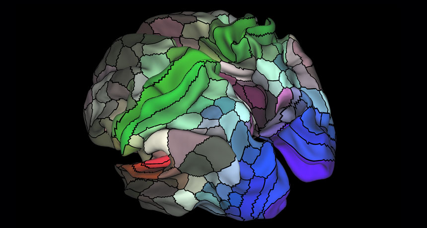
Analyzing a bevy of diverse data, scientists have drawn a new map of the human brain in extreme relief. Their approach demarcated 180 areas in each half of the outer layer of the brain — including 97 regions in each half that haven’t been described before. The high-resolution map will allow scientists to more precisely scrutinize brain regions and see how they change with, for instance, age and disease.
Many previous maps of the brain have been built with just one type of data. The new map, described July 20 in Nature, forms a holistic view of the brain by combining several different types of information. These specs included how areas behaved while doing certain tasks or nothing at all, as well as detailed anatomical data about the shape and thickness of the brain. Using these metrics from 210 healthy people, neuroscientist David Van Essen of Washington University in St. Louis and colleagues found that each hemisphere contains 180 distinct areas (separated by black lines in image). In this view, colors show how tightly linked each area is to other brain areas that handle auditory (red), touch and movement (green) or visual (blue) information.
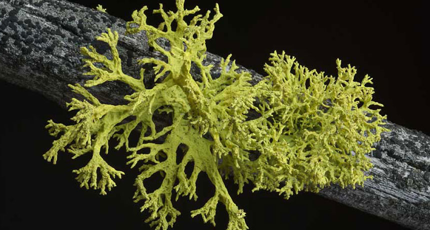
The discovery of unknown yeasts hiding in lichens from six continents could shake up a basic idea of what makes up a lichen partnership.
For more than a century, biologists have described a lichen as a fungus growing intimately with some microbes (algae and/or cyanobacteria) that harvest solar energy. The fungus is treated as so important that its name serves as the name for the whole lichen.
Biologists have recognized that more than one fungus can show up in lichen close-ups, but their role hasn’t been clear. Now that may be on the brink of changing. Fifty-two genera of lichens collected from around the world include a second fungus — single cells, called yeasts, of a previously unknown order now christened Cyphobasidiales. Toby Spribille of the University of Graz in Austria and colleagues report the finding online July 21 in Science. The first example discovered illustrates why these yeasts might turn out to be more than parasites or mere hitchhikers, says study coauthor John McCutcheon of the University of Montana in Missoula. He and Spribille started the research out of curiosity. They wondered how the yellow, toxin-bearing, thready tangles of lichen called Bryoria tortuosa could have the same fungus and the same algal partner — and thus technically be the same species — as the brown, toxin-poor lichen traditionally called B. fremontii. The researchers looked to see which genes were active in each lichen in hopes that some discrepancy could explain the difference in forms. What the researchers found had nothing to do with the alga or previously known fungus. Instead, ample genetic activity of more abundant yeasts in the toxic B. tortuosa turned out to be the most striking disparity.
After five years of work, the research team now has microscope images of yeast cells embedded in the outer layer, or cortex, of B. tortuosa. Gene-activity results suggest that the yeasts could be what’s making the difference between the forms, maybe even synthesizing toxic vulpinic acid. The yeasts turning up across this widespread class of lichens might explain other mysteries, such as why researchers have largely failed to re-create lichen partnerships in the lab. It’s a bold hypothesis, but lichenologist Robert Lücking of the Botanic Garden and Botanical Museum Berlin-Dahlem takes the idea of yeast partners seriously. “This will be a huge surprise to the lichenological and mycological community,” he says.
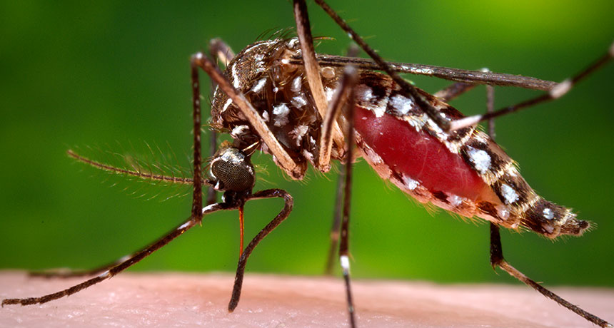
Mosquitoes in Miami now appear to be transmitting Zika virus.
Four cases of Zika infection in Florida were probably acquired via the bite of local mosquitoes, the state’s health department announced July 29. These are the first cases of local transmission of the virus in the continental United States.
“Zika is now here,” Tom Frieden, director of U.S. Centers for Disease Control and Prevention, said in a news briefing July 29.
No mosquitoes trapped yet have tested positive for the virus, but officials suspect Aedes aegypti mosquitoes in a several-block area in north Miami are to blame. “Everything we’ve seen so far indicates that this is mosquito-borne transmission,” Frieden said.
Florida’s small cluster of cases does not necessarily foreshadow an epidemic, he said. The four infected people probably were bitten in early July. Since then, Florida has stepped up efforts to stamp out mosquitoes — including going door-to-door to get rid of standing water and spraying insecticides by truck and by people on foot.
“We believe that widespread transmission in the continental U.S. is unlikely,” Frieden said. “But it’s not impossible.”
Two other mosquito-borne diseases, dengue and chikungunya, have spread locally in Florida in the past. But, Frieden said, those diseases generally dead-end after infecting just one person.
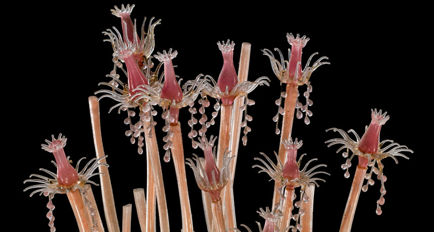
From 1863 to 1890, Leopold and Rudolf Blaschka made more than 10,000 sea creatures out of glass. There were anemones with tapered tentacles and pearled undersides, translucent jellyfish trailing the most delicate threads and feather stars more than worthy of their name despite their rigid composition. The intricate invertebrates, crafted by the father-son team at their studio in Dresden, Germany, were shipped across the world to serve as teaching models at universities and museums. In an era before marine surveys and underwater photography, before the rise of scuba diving resorts, the Blaschkas showed the world the wonders of the sea.
Over five dozen of their glass wonders are now on display at the Corning Museum of Glass in “Fragile Legacy.” Though the exhibit opens with glass eyeballs and a piece of jewelry — a nod to the Blaschkas’ pre-invertebrate business — the highlight is a darkened room set up like an aquarium, with sea creatures seemingly floating in blue. There’s a notable absence of museum placards and descriptions. “We really want people to look at the glassiness,” says Marvin Bolt, a curator of the exhibit, before pointing out the “Field Guide to Underwater Models.” The pamphlet contains each animal’s species name, as it was known in 1885 (when Cornell University acquired the pieces, now on loan to Corning) and as it is known today. The aquarium offers a sense of the Blaschkas’ style, but it’s the room next door that provides the substance. Sketches and watercolors, bottles of colored powders, tweezers, pliers, scoops and wire, along with a demonstration video, give a fuller sense of how the Blaschkas did their work. Equally impressive are the matchboxes filled with kleine augen (“little eyes” in German) and other tiny but uniform component pieces, suggestive of an assembly line approach to handcrafting the final glass forms. A series of case studies explains how conservators stabilized the pieces, and a trailer for a related documentary, also titled Fragile Legacy, highlights the vulnerability, not of the glass, but of the real-world creatures living in warming seas. There’s one thing you won’t find in this exhibit — the flowers that the Blaschkas are most famous for today, commissioned by Harvard beginning in 1886. But you’ll spot seeds of this later work in the sea animals’ slender stalks and garlands of orbs. As the Blaschkas moved on to new subjects, their artistry evolved from the forms they’d already mastered.
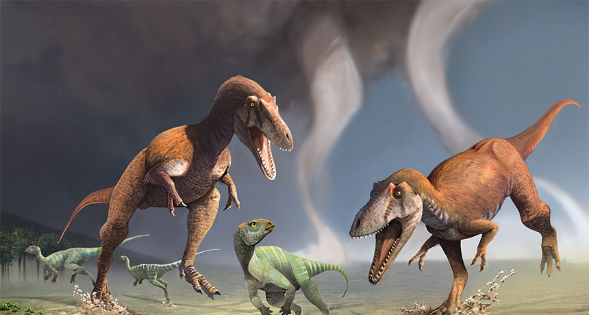
What had two puny arms, lived 90 million years ago and probably chowed down on other dinosaurs? (Hint: It’s not T. rex.)
A new dinosaur discovered in what is now Patagonia had the runty forelimbs of a Tyrannosaurus rex but is no cousin of the giant iconic predator, researchers report July 13 in PLOS ONE.
The new species, Gualicho shinyae, has a close relative in Africa, an analysis of fossils suggests. T. rex’s ancestors, on the other hand, came from Asia. Gualicho is a “smaller, slimmer, trimmer version of a T. rex,” says study coauthor Peter Makovicky, a paleontologist at the Field Museum in Chicago. It probably weighed about a ton and was longer than a pickup truck. In 2007, Makovicky’s team discovered Gualicho’s partial skeleton — including those impractical arms. The dinosaur probably caught prey with its huge head, Makovicky says. Though the researchers haven’t dug up a skull yet, tiny arms seem to be a trade-off for a big head. Finding Gualicho’s skull would help nail down that idea, he adds.
Gualicho may have fed on grazers called ornithopods, such as duck-billed dinosaurs. Or perhaps it fed on the long-necked, long-tailed sauropods, which were common in the region. But only the youngsters. Sauropod adults were gigantic, Makovicky says, definitely not prey for a (relatively) little guy like Gualicho.
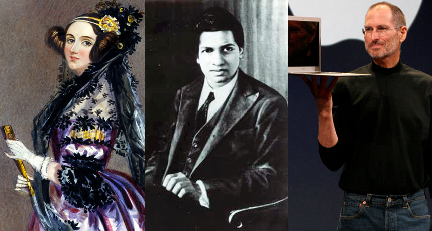
It’s hard to pin down Stephen Wolfram’s scientific discipline. He is part mathematician, part computer scientist, part physicist. He’s also an inventor and entrepreneur, known for the mathematics software package Mathematica and a variety of other endeavors. And he blogs.
Among his blog posts and other essays and talks are commentaries on the lives of other prominent figures from science and math; some Wolfram knew personally, others he has researched extensively. In his new book Idea Makers, Wolfram has collected accounts of 16 such people, discussing their work and its meaning for the nature of science and the process of understanding math, computing and the physical world. Each entry provides a healthy dose of personal information as well. Some of the people Wolfram discusses are widely known — Richard Feynman and Steve Jobs, for example. Others are relatively obscure, such as Russell Towle and Richard Crandall. But all have captivated Wolfram’s interest, either by way of friendship or their historical importance for the fields of study that Wolfram himself has contributed to.
On the historical side, Wolfram offers his views of the logician Kurt Gödel, computer scientist (perhaps the original computer scientist) Alan Turing and mathematicians John von Neumann and George Boole. Wolfram provides an especially extensive discussion of Ada Lovelace and her interactions with Charles Babbage as they contemplated the prospect of powerful computing engines a century ahead of their time.
Wolfram also dives into the story of Srinivasa Ramanujan, and the lessons his genius offers about the nature of math. With little formal training, Ramanujan discovered many surprising results that seemed at first glance to be a bunch of “random facts of mathematics.” But in recent decades, many have been linked to deep mathematical principles that he seems to have somehow perceived without knowing it. How did he do it? Wolfram suspects that he “had intuition and aesthetic criteria that in some sense captured some of the deeper principles we now know, even if he couldn’t express them directly.”
Personal style, whether as reflected in the subtle genius of Ramanujan or the boldness of vision-driven Jobs, plays an underappreciated role in the progress of science and technology. Wolfram has collected some illuminating examples of the ways the human side of scientific thinkers can enrich the work they do.
The virus, which can cause brain damage in infants infected in the womb, kills stem cells and stunts their numbers in the brains of adult mice, researchers report August 18 in Cell Stem Cell. Though scientists have considered Zika primarily a threat to unborn babies, the new findings suggest that the virus may cause unknown — and potentially long-term — damage to adults as well.
In adults, Zika has been linked to Guillain-Barré syndrome, a rare neurological disorder (SN: 4/2/16, p. 29). But for most people, infection is typically mild: a headache, fever and rash lasting up to a week, or no symptoms at all. In pregnant women, though, the virus can lodge in the brain of a fetus and kill off newly developing cells (SN: 4/13/16). If Zika targets newborn brain cells, adults may be at risk, too, reasoned neuroscientist Joseph Gleeson of Rockefeller University in New York City and colleagues. Parts of the forebrain and the hippocampus, which plays a crucial role in learning and memory, continue to generate nerve cells in adult brains.
In mice infected with Zika, the virus hit these brain regions hard. Nerve cells died and the regions generated one-fifth to one-half as many new cells compared with those of uninfected mice. The results might not translate to humans; the mice were genetically engineered to have weak immune systems, making them susceptible to Zika.
But Zika could potentially harm immunocompromised people and perhaps even healthy people in a similar way, the authors write.
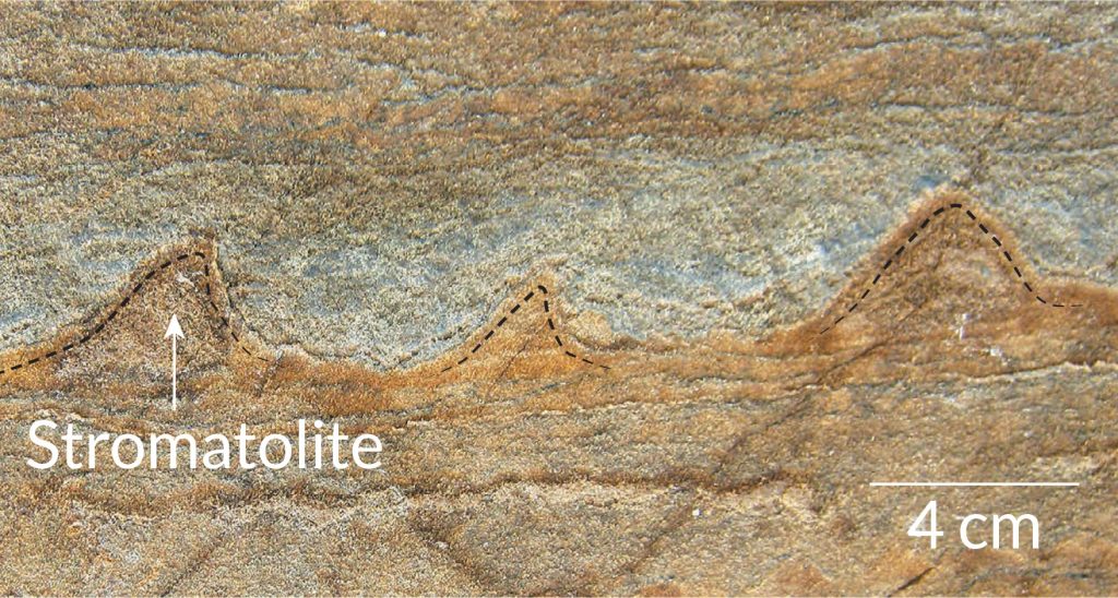
A melting snow patch in Greenland has revealed what could be the oldest fossilized evidence of life on Earth. The 3.7-billion-year-old structures may help scientists retrace the rise of the first organisms relatively soon after Earth’s formation around 4.5 billion years ago (SN: 2/8/14, p. 16), the discoverers report online August 31 in Nature.
Unlike dinosaur bones, the new fossils are not preserved bits of an ancient critter. The Greenland fossils are mounds of minerals a few centimeters tall that may have been deposited by clusters of microbes several hundred million years after Earth formed. The shape and chemical composition of the mounds, called stromatolites, match those formed by modern bacterial communities living in shallow seawater, says a team led by geologist Allen Nutman of the University of Wollongong in Australia.
If confirmed, the fossils demonstrate that sophisticated, mound-building microbial life appeared early on in Earth’s history. That early start backs up previous genetic and chemical studies that place the advent of basic life on Earth before 4 billion years ago (SN Online: 10/19/15).
Aneil Agrawal, his rangy frame at ease on a black metal street bench, is staring into some midair memory and speaking about disgust.
“I was first exposed to the idea of theoretical biology as an undergraduate and I actually hated it,” he says. “I loved biology and I liked math — it was like two different food types that you like but the two of them together are going to be terrible.”
Since then, he has remained a fan of the two foods, and his distaste for combining them has turned into enthusiasm strong enough to build a career on. Agrawal, now a 41-year-old evolutionary geneticist at the University of Toronto, both builds mathematical descriptions of biological processes and leads what he describes as “insanely laborious” experiments with fruit flies, duckweed and microscopic aquatic animals called rotifers. Often experimentalists venturing into theory “dabble and do some stuff, but it’s not very good,” says evolutionary biologist Mark Kirkpatrick of the University of Texas at Austin. Agrawal, however, is “one of the few people who’s doing really good theory and really good experimental work.”
Two of the themes Agrawal works on — the evolution of sex and the buildup over time of harmful mutations — are “very deep and important problems in evolutionary biology,” Kirkpatrick says. Agrawal and colleagues have made a case for a once-fringe idea: that an abundance of harmful mutations can invite even more harmful mutations. Agrawal’s work has also provided rare data to support the idea that the need to adapt to new circumstances has favored sexual over asexual reproduction. Why sexual reproduction is much more common among complex life-forms has been a long-standing puzzle in biology. Life’s complexity appealed to Agrawal from childhood; he remembers days playing among the backyard bugs and frogs in suburban Vancouver. At first, he imagined his grown-up life out in the field, “living in a David Attenborough show.” As he grew older though, he discovered he was a lab animal: “I was more interested in being able to ask more precise questions under more controlled circumstances.”
Sally Otto, now president-elect of the Society for the Study of Evolution, met Agrawal in the 1990s when he was an undergraduate at the University of British Columbia in Vancouver. He returned to Vancouver in 2003, after earning his Ph.D., to do experimental work and “beef up his ability to do theory,” she says. She cosupervised his postdoctoral effort. Agrawal “picks up theory very quickly,” Otto says. Knowing a huge amount of math to begin with is less important than having insight into what math to learn. The first alluring ideas about how to approach a puzzle often don’t work out, she says, so “there’s a certain doggedness — you have to really keep at it.”
Agrawal needed some time before he came around to theoretical biology. It disgusted him, he says, because he expected it to take the rich variety out of biology. “The reason many people, including me, were attracted to biology was because it’s not boxes and triangles,” he says. “It’s complicated and interesting.” At first he thought modeling a biological process mathematically “sterilized it.” But he eventually found that mathematical description could “help to clarify our thinking about the wonderful mess of diversity that’s out there.” At the street bench, Agrawal muses about how he tends to “think quantitatively.” His father has a Ph.D. in engineering, but “we weren’t the kind of family that had to do math problems at the dinner table.” He laughs. “Though I do that to my own kids.” His success so far is mixed, depending in part on whether he catches his two sons, ages 10 and 7, in the right mood. Agrawal also thinks intensely, possibly another secret to his success — he has received more than half a dozen awards and prizes, including the 2015 Steacie Prize for Natural Sciences. The bench where we’ve settled is only half a block from the conference center in Austin, where Evolution 2016, the field’s biggest meeting of the year, has hit day four of its five-day marathon. Agrawal gave one of the first talks, a smooth, perfectly timed zoom through a recent fly experiment. He is a coauthor on five more presentations, along with chairing one of the frenetic sessions where talks are compressed into five minutes. By this point, many of the 1,800 or so attendees are showing strain — wearing name tags wrong side frontward, snoring open-mouthed in hallway chairs or flailing their arms in conversations fueled by way too much caffeine. Agrawal, however, seems relaxed, listening quietly, staring off in thought, speaking in quiet bursts. This guy can focus.
One of his early theory papers studied mutation accumulation. Previous work had suggested that microbes in stressful environments, compared with microbes lapped in luxury, are more likely to make mistakes in copying genes that then get passed on to the next generation. Agrawal wondered whether cells that are stressed for another reason — an already heavy burden of harmful mutations — would likewise be more inclined to build up additional mutations. He calls this scenario “a spiral of doom.”
The idea intrigued him because he suspected that sexual reproduction would do a better job of purging these mutations than asexual reproduction. “What I found in doing the theory was that I was exactly wrong,” he says. The sexual populations would end up with more, not fewer, mutations.
Though the theory part of the paper turned out well, the journal Genetics rejected it — there was hardly any experimental evidence that the scenario would arise in the real world.
Agrawal published the paper elsewhere in 2002 and, when he began setting up his own lab at the University of Toronto, he returned to the idea. In the years since, he and colleagues have published a string of papers adding evidence to the argument. They have found, for example, that fruit flies burdened with misbegotten genes lag in growth and struggle to keep their DNA in good repair. The idea is no longer airy speculation, says Charles Baer, who’s checking for mutation accumulation in nematodes at the University of Florida in Gainesville.
Chrissy Spencer, a postdoc during the early years of Agrawal’s mutation studies, points out that a vital skill of a good experimentalist is just knowing intuitively whether a species is right for a certain kind of test. Agrawal has that knack, for better and for worse. For some studies on the evolution of sex, Agrawal eventually turned to rotifers. The stubby little cylinders with a circlet of hairy projections around their mouths can reproduce either sexually or asexually, so they’re great for testing what factors favor one over the other. Rotifers, however, are also “finicky,” he says. His students have cared for them, sometimes for months, only to have them all die for no discernible reason, sometimes before generating any data.
Having the practitioner’s inside view of experiments and theory may help Agrawal, but it also has its costs. “There are better theoreticians out there and there are better experimentalists,” he says, and he wishes at times that he was more solidly in one camp or the other. He pauses and then, a biologist to the core, says: “That’s my niche.”
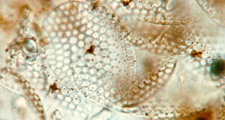
DENVER — Life on Earth got into the shell game more than 200 million years earlier than previously thought.
Fossilized eukaryotes — complex life-forms that include animals and plants — discovered in Canada are decked out in armorlike layers of mineral plates, paleobiologist Phoebe Cohen said September 27 at the Geological Society of America’s annual meeting. At about 809 million years old, the find is the oldest evidence of organisms controlling the formation of minerals, a process called biomineralization. This new origin of biomineralization coincides with major changes that mark the end of a period known as the “boring billion” (SN: 11/14/15, p. 18), said Stanford University paleontologist Erik Sperling, who was not involved in the discovery. “There were big things going on with ocean chemistry,” he said. “It’s interesting to see the biological response.”
These ancient eukaryotes built their exoskeletons using a very different process from most modern shell-making microbes. That uniqueness offers insights into how the mineral-making ability first evolved, said Cohen, who studies ancient ecosystems at Williams College in Williamstown, Mass.
“We have been able to identify specific conditions that facilitated the evolution of the first eukaryote to biomineralize in Earth’s history,” she said. “It paints a beautiful picture of the ecology and evolution and environmental conditions that led to this dramatic innovation.”
Donning an exoskeleton of minerals protects microbes from predators and forms a crucial stage in the modern carbon cycle. The shells make marine microbes such as certain phytoplankton species sink faster after they die, removing carbon from the upper ocean. Previous clear evidence of eukaryote biomineralization dates back to around 560 million years ago in early corallike animals.
Odd fossils discovered in the late 1970s and covered in mineral plates shaped like circles, squares and “Honeycomb cereal” (as Cohen described them) hinted that the skill evolved much earlier, but the discovery raised many questions. Dating techniques then put the age of the fossils somewhere within a 100-million-year range from about 811 million to 717 million years ago, and scientists couldn’t rule out that the fossils’ scalelike minerals formed after the organisms died. Cohen and colleagues revisited these curious fossils. By accurately dating the organic-rich shale a few meters below the fossils in the rock record, the researchers pegged the fossils’ age at 809 million years old, give or take a few million years. An electron microscope let researchers see that each plate is a weave of elongated mineral fibers. This intricate, orderly design had to have been purposefully built by life manipulating mineral formation, Cohen said.
The mineral plates themselves are odd. Most modern microbes make shells out of calcium carbonate, but the ancient shells are made of calcium phosphate, the same crystal used in human teeth enamel. Today, phosphate is limited in the environment and most microbes avoid wasting it.
That may not have been as much of an issue in the marine basin where the eukaryotes lived, the researchers found. Analysis of rocks surrounding the fossils indicate that the amount of oxygen in the waters where the eukaryotes lived was inconsistent. Fluctuating oxygen levels pulled phosphate from underlying sediment into the water, where it was available for mineral making. These favorable conditions plus the need for protection from predation (SN: 11/28/15, p. 13) probably drove the first evolution of biomineralization, Cohen said. Eventually the environment changed, and these shell-making species died out.