It’s hard to pin down Stephen Wolfram’s scientific discipline. He is part mathematician, part computer scientist, part physicist. He’s also an inventor and entrepreneur, known for the mathematics software package Mathematica and a variety of other endeavors. And he blogs.
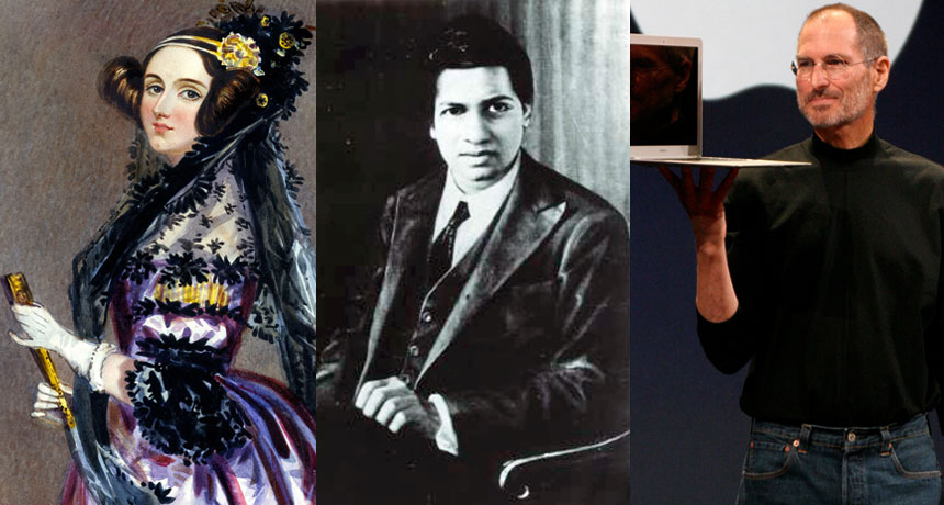
Among his blog posts and other essays and talks are commentaries on the lives of other prominent figures from science and math; some Wolfram knew personally, others he has researched extensively. In his new book Idea Makers, Wolfram has collected accounts of 16 such people, discussing their work and its meaning for the nature of science and the process of understanding math, computing and the physical world. Each entry provides a healthy dose of personal information as well. Some of the people Wolfram discusses are widely known — Richard Feynman and Steve Jobs, for example. Others are relatively obscure, such as Russell Towle and Richard Crandall. But all have captivated Wolfram’s interest, either by way of friendship or their historical importance for the fields of study that Wolfram himself has contributed to.
On the historical side, Wolfram offers his views of the logician Kurt Gödel, computer scientist (perhaps the original computer scientist) Alan Turing and mathematicians John von Neumann and George Boole. Wolfram provides an especially extensive discussion of Ada Lovelace and her interactions with Charles Babbage as they contemplated the prospect of powerful computing engines a century ahead of their time.
Wolfram also dives into the story of Srinivasa Ramanujan, and the lessons his genius offers about the nature of math. With little formal training, Ramanujan discovered many surprising results that seemed at first glance to be a bunch of “random facts of mathematics.” But in recent decades, many have been linked to deep mathematical principles that he seems to have somehow perceived without knowing it. How did he do it? Wolfram suspects that he “had intuition and aesthetic criteria that in some sense captured some of the deeper principles we now know, even if he couldn’t express them directly.”
Personal style, whether as reflected in the subtle genius of Ramanujan or the boldness of vision-driven Jobs, plays an underappreciated role in the progress of science and technology. Wolfram has collected some illuminating examples of the ways the human side of scientific thinkers can enrich the work they do.
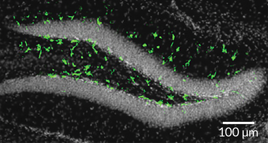
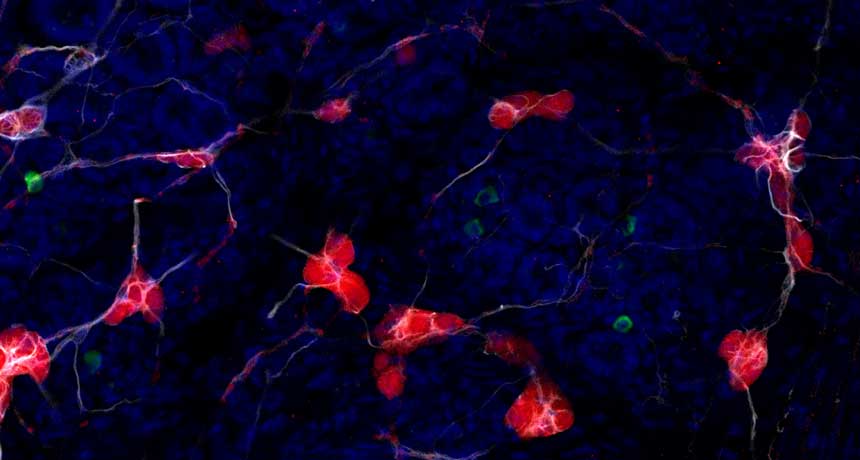
The virus, which can cause brain damage in infants infected in the womb, kills stem cells and stunts their numbers in the brains of adult mice, researchers report August 18 in Cell Stem Cell. Though scientists have considered Zika primarily a threat to unborn babies, the new findings suggest that the virus may cause unknown — and potentially long-term — damage to adults as well.
In adults, Zika has been linked to Guillain-Barré syndrome, a rare neurological disorder (SN: 4/2/16, p. 29). But for most people, infection is typically mild: a headache, fever and rash lasting up to a week, or no symptoms at all. In pregnant women, though, the virus can lodge in the brain of a fetus and kill off newly developing cells (SN: 4/13/16). If Zika targets newborn brain cells, adults may be at risk, too, reasoned neuroscientist Joseph Gleeson of Rockefeller University in New York City and colleagues. Parts of the forebrain and the hippocampus, which plays a crucial role in learning and memory, continue to generate nerve cells in adult brains.
In mice infected with Zika, the virus hit these brain regions hard. Nerve cells died and the regions generated one-fifth to one-half as many new cells compared with those of uninfected mice. The results might not translate to humans; the mice were genetically engineered to have weak immune systems, making them susceptible to Zika.
But Zika could potentially harm immunocompromised people and perhaps even healthy people in a similar way, the authors write.
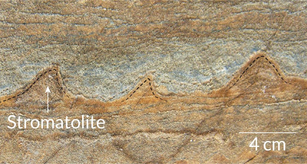
A melting snow patch in Greenland has revealed what could be the oldest fossilized evidence of life on Earth. The 3.7-billion-year-old structures may help scientists retrace the rise of the first organisms relatively soon after Earth’s formation around 4.5 billion years ago (SN: 2/8/14, p. 16), the discoverers report online August 31 in Nature.
Unlike dinosaur bones, the new fossils are not preserved bits of an ancient critter. The Greenland fossils are mounds of minerals a few centimeters tall that may have been deposited by clusters of microbes several hundred million years after Earth formed. The shape and chemical composition of the mounds, called stromatolites, match those formed by modern bacterial communities living in shallow seawater, says a team led by geologist Allen Nutman of the University of Wollongong in Australia.
If confirmed, the fossils demonstrate that sophisticated, mound-building microbial life appeared early on in Earth’s history. That early start backs up previous genetic and chemical studies that place the advent of basic life on Earth before 4 billion years ago (SN Online: 10/19/15).
Aneil Agrawal, his rangy frame at ease on a black metal street bench, is staring into some midair memory and speaking about disgust.
“I was first exposed to the idea of theoretical biology as an undergraduate and I actually hated it,” he says. “I loved biology and I liked math — it was like two different food types that you like but the two of them together are going to be terrible.”
Since then, he has remained a fan of the two foods, and his distaste for combining them has turned into enthusiasm strong enough to build a career on. Agrawal, now a 41-year-old evolutionary geneticist at the University of Toronto, both builds mathematical descriptions of biological processes and leads what he describes as “insanely laborious” experiments with fruit flies, duckweed and microscopic aquatic animals called rotifers. Often experimentalists venturing into theory “dabble and do some stuff, but it’s not very good,” says evolutionary biologist Mark Kirkpatrick of the University of Texas at Austin. Agrawal, however, is “one of the few people who’s doing really good theory and really good experimental work.”
Two of the themes Agrawal works on — the evolution of sex and the buildup over time of harmful mutations — are “very deep and important problems in evolutionary biology,” Kirkpatrick says. Agrawal and colleagues have made a case for a once-fringe idea: that an abundance of harmful mutations can invite even more harmful mutations. Agrawal’s work has also provided rare data to support the idea that the need to adapt to new circumstances has favored sexual over asexual reproduction. Why sexual reproduction is much more common among complex life-forms has been a long-standing puzzle in biology. Life’s complexity appealed to Agrawal from childhood; he remembers days playing among the backyard bugs and frogs in suburban Vancouver. At first, he imagined his grown-up life out in the field, “living in a David Attenborough show.” As he grew older though, he discovered he was a lab animal: “I was more interested in being able to ask more precise questions under more controlled circumstances.”
Sally Otto, now president-elect of the Society for the Study of Evolution, met Agrawal in the 1990s when he was an undergraduate at the University of British Columbia in Vancouver. He returned to Vancouver in 2003, after earning his Ph.D., to do experimental work and “beef up his ability to do theory,” she says. She cosupervised his postdoctoral effort. Agrawal “picks up theory very quickly,” Otto says. Knowing a huge amount of math to begin with is less important than having insight into what math to learn. The first alluring ideas about how to approach a puzzle often don’t work out, she says, so “there’s a certain doggedness — you have to really keep at it.”
Agrawal needed some time before he came around to theoretical biology. It disgusted him, he says, because he expected it to take the rich variety out of biology. “The reason many people, including me, were attracted to biology was because it’s not boxes and triangles,” he says. “It’s complicated and interesting.” At first he thought modeling a biological process mathematically “sterilized it.” But he eventually found that mathematical description could “help to clarify our thinking about the wonderful mess of diversity that’s out there.” At the street bench, Agrawal muses about how he tends to “think quantitatively.” His father has a Ph.D. in engineering, but “we weren’t the kind of family that had to do math problems at the dinner table.” He laughs. “Though I do that to my own kids.” His success so far is mixed, depending in part on whether he catches his two sons, ages 10 and 7, in the right mood. Agrawal also thinks intensely, possibly another secret to his success — he has received more than half a dozen awards and prizes, including the 2015 Steacie Prize for Natural Sciences. The bench where we’ve settled is only half a block from the conference center in Austin, where Evolution 2016, the field’s biggest meeting of the year, has hit day four of its five-day marathon. Agrawal gave one of the first talks, a smooth, perfectly timed zoom through a recent fly experiment. He is a coauthor on five more presentations, along with chairing one of the frenetic sessions where talks are compressed into five minutes. By this point, many of the 1,800 or so attendees are showing strain — wearing name tags wrong side frontward, snoring open-mouthed in hallway chairs or flailing their arms in conversations fueled by way too much caffeine. Agrawal, however, seems relaxed, listening quietly, staring off in thought, speaking in quiet bursts. This guy can focus.
One of his early theory papers studied mutation accumulation. Previous work had suggested that microbes in stressful environments, compared with microbes lapped in luxury, are more likely to make mistakes in copying genes that then get passed on to the next generation. Agrawal wondered whether cells that are stressed for another reason — an already heavy burden of harmful mutations — would likewise be more inclined to build up additional mutations. He calls this scenario “a spiral of doom.”
The idea intrigued him because he suspected that sexual reproduction would do a better job of purging these mutations than asexual reproduction. “What I found in doing the theory was that I was exactly wrong,” he says. The sexual populations would end up with more, not fewer, mutations.
Though the theory part of the paper turned out well, the journal Genetics rejected it — there was hardly any experimental evidence that the scenario would arise in the real world.
Agrawal published the paper elsewhere in 2002 and, when he began setting up his own lab at the University of Toronto, he returned to the idea. In the years since, he and colleagues have published a string of papers adding evidence to the argument. They have found, for example, that fruit flies burdened with misbegotten genes lag in growth and struggle to keep their DNA in good repair. The idea is no longer airy speculation, says Charles Baer, who’s checking for mutation accumulation in nematodes at the University of Florida in Gainesville.
Chrissy Spencer, a postdoc during the early years of Agrawal’s mutation studies, points out that a vital skill of a good experimentalist is just knowing intuitively whether a species is right for a certain kind of test. Agrawal has that knack, for better and for worse. For some studies on the evolution of sex, Agrawal eventually turned to rotifers. The stubby little cylinders with a circlet of hairy projections around their mouths can reproduce either sexually or asexually, so they’re great for testing what factors favor one over the other. Rotifers, however, are also “finicky,” he says. His students have cared for them, sometimes for months, only to have them all die for no discernible reason, sometimes before generating any data.
Having the practitioner’s inside view of experiments and theory may help Agrawal, but it also has its costs. “There are better theoreticians out there and there are better experimentalists,” he says, and he wishes at times that he was more solidly in one camp or the other. He pauses and then, a biologist to the core, says: “That’s my niche.”
DENVER — Life on Earth got into the shell game more than 200 million years earlier than previously thought.
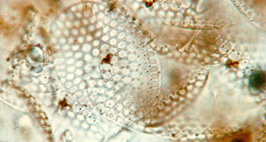
Fossilized eukaryotes — complex life-forms that include animals and plants — discovered in Canada are decked out in armorlike layers of mineral plates, paleobiologist Phoebe Cohen said September 27 at the Geological Society of America’s annual meeting. At about 809 million years old, the find is the oldest evidence of organisms controlling the formation of minerals, a process called biomineralization. This new origin of biomineralization coincides with major changes that mark the end of a period known as the “boring billion” (SN: 11/14/15, p. 18), said Stanford University paleontologist Erik Sperling, who was not involved in the discovery. “There were big things going on with ocean chemistry,” he said. “It’s interesting to see the biological response.”
These ancient eukaryotes built their exoskeletons using a very different process from most modern shell-making microbes. That uniqueness offers insights into how the mineral-making ability first evolved, said Cohen, who studies ancient ecosystems at Williams College in Williamstown, Mass.
“We have been able to identify specific conditions that facilitated the evolution of the first eukaryote to biomineralize in Earth’s history,” she said. “It paints a beautiful picture of the ecology and evolution and environmental conditions that led to this dramatic innovation.”
Donning an exoskeleton of minerals protects microbes from predators and forms a crucial stage in the modern carbon cycle. The shells make marine microbes such as certain phytoplankton species sink faster after they die, removing carbon from the upper ocean. Previous clear evidence of eukaryote biomineralization dates back to around 560 million years ago in early corallike animals.
Odd fossils discovered in the late 1970s and covered in mineral plates shaped like circles, squares and “Honeycomb cereal” (as Cohen described them) hinted that the skill evolved much earlier, but the discovery raised many questions. Dating techniques then put the age of the fossils somewhere within a 100-million-year range from about 811 million to 717 million years ago, and scientists couldn’t rule out that the fossils’ scalelike minerals formed after the organisms died. Cohen and colleagues revisited these curious fossils. By accurately dating the organic-rich shale a few meters below the fossils in the rock record, the researchers pegged the fossils’ age at 809 million years old, give or take a few million years. An electron microscope let researchers see that each plate is a weave of elongated mineral fibers. This intricate, orderly design had to have been purposefully built by life manipulating mineral formation, Cohen said.
The mineral plates themselves are odd. Most modern microbes make shells out of calcium carbonate, but the ancient shells are made of calcium phosphate, the same crystal used in human teeth enamel. Today, phosphate is limited in the environment and most microbes avoid wasting it.
That may not have been as much of an issue in the marine basin where the eukaryotes lived, the researchers found. Analysis of rocks surrounding the fossils indicate that the amount of oxygen in the waters where the eukaryotes lived was inconsistent. Fluctuating oxygen levels pulled phosphate from underlying sediment into the water, where it was available for mineral making. These favorable conditions plus the need for protection from predation (SN: 11/28/15, p. 13) probably drove the first evolution of biomineralization, Cohen said. Eventually the environment changed, and these shell-making species died out.
ORLANDO, Fla. — Here’s another reason not to love car exhaust: The fumes may make it harder for honeybees to learn floral scents.
In lab tests, bees normally caught on quickly that a puff of floral scent meant a researcher would soon offer them a taste of sugar, Ryan James Leonard of the University of Sydney said September 30 at the International Congress of Entomology. After two sequences of puff-then-sugar, just a whiff of fragrance typically made the bees stick out their tongues. But when that floral scent was mixed with vehicle exhaust, it took the bees several more run-throughs to respond to the puff signal. Honeybees buzzing among roadside flowers must contend with vehicle pollution as they learn various foraging cues. Another lab reported in 2013 that diesel exhaust reacted with some of the chemical components of canola flowers, rendering them more difficult for bees to recognize.
Building on that concern, Leonard and colleagues found that it was easy for bees to learn the scent of linalool, a widespread ingredient in many flower fragrances, whether mixed with exhaust fumes or not. But exhaust made it take longer than two trials for bees to learn the scent ingredients myrcene (three trials), dipentene (four) and the full, multicomponent fragrance of geraniums (six).
Road ecologists have put a lot of effort into studying how vehicles kill animals. But Leonard hopes for more interest now in how chronic exposure to traffic affects living animals.
When small lies snowball into blizzards of deception, the brain becomes numb to dishonesty. As people tell more and bigger lies, certain brain areas respond less to the whoppers, scientists report online October 24 in Nature Neuroscience. The results might help explain how small transgressions can ultimately set pants aflame.
The findings “have big implications for how lying can develop,” says developmental psychologist Victoria Talwar of McGill University in Montreal, who studies how dishonest behavior develops in children. “It starts to give us some idea about how lying escalates from small lies to bigger ones.” During the experiment, researchers from University College London and Duke University showed 80 participants a crisp, big picture of a glass jar of pennies. They were told that they needed to send an estimate of how much money was in the jar to an unseen partner who saw a smaller picture of the same jar. Each participant was serving as a “well-informed financial adviser tasked with advising a client who is less informed about what investments to make,” study coauthor Neil Garrett of University College London said October 20 during a news briefing. Researchers gave people varying incentives to lie. In some cases, for instance, intentionally overestimating the jar’s contents was rewarded with a bigger cut of the money.
As the experiment wore on, the fibs started flying. People lied the most when the lie would benefit both themselves and their unseen partner. But these “financial advisers” still told self-serving lies even when it would hurt their partner.
Twenty-five participants underwent fMRI scans while lying. When a person had previously lied, brain activity lessened in certain areas of the brain, most notably in the amygdala. A pair of almond-shaped brain structures nestled deep in the brain, the amygdalae are tightly linked to emotions. This diminished amygdala activity could even predict whether a person would lie on the next trial, results that suggest that the reduced brain activity is actually influencing the decision to lie.
The study design gets around a problem that confounds other lying experiments, says neuroscientist Bernd Weber of the University of Bonn in Germany. Many experiments are based on lies that people have been instructed to say, a situation that “hardly resembles real-world behavior,” he says. Here, the participants were self-motivated liars.
Without any negative consequences from their lies, participants weren’t afraid of being caught. That impunity might affect activity in the amygdala, Weber says. Further experiments are needed to reveal the effects of such fear. From Ponzi schemes to current politics, case studies abound of small lies spiraling into much bigger deceits, study coauthor Tali Sharot of the University College London said in the news briefing. “There are many reasons why this might happen, societal reasons, but we suspected that there might be a basic biological principle of how our brain works that contributes to this phenomenon,” she said.
The principle she had in mind is called emotional adaptation — the same phenomenon that explains why the scent of strong perfume becomes less noticeable over time. The first time you cheat on your taxes, you’d probably feel quite bad about it, Sharot said. That bad feeling is good, because it curbs your dishonesty. “The next time you cheat, you have already adapted,” she said. “There’s less negative reaction to hold you back so you might be lying more.”
Narwhals use highly targeted beams of sound to scan their environment for threats and food. In fact, the so-called unicorns of the sea (for their iconic head tusks) may produce the most refined sonar of any living animal.
A team of researchers set up 16 underwater microphones to eavesdrop on narwhal click vocalizations at 11 ice pack sites in Greenland’s Baffin Bay in 2013. The recordings show that narwhal clicks are extremely intense and directional — meaning they can widen and narrow the beam of sound to find prey over long and short distances. It’s the most directional sonar signal measured in a living species, the researchers report November 9 in PLOS ONE.
The sound beams are also asymmetrically narrow on top. That minimizes clutter from echoes bouncing off the sea surface or ice pack. Finally, narwhals scan vertically as they dive, which could help them find patches of open water where they can surface and breathe amid sea ice cover. All this means that narwhals employ pretty sophisticated sonar.
The audio data could help researchers tell the difference between narwhal vocalizations and those of neighboring beluga whales. It also provides a baseline for assessing the potential impact of noise pollution from increases in shipping traffic made possible by sea ice loss.
SAN DIEGO — Over the course of months, clumps of a protein implicated in Parkinson’s disease can travel from the gut into the brains of mice, scientists have found.
The results, reported November 14 at the annual meeting of the Society for Neuroscience, suggest that in some cases, Parkinson’s may get its start in the gut. That’s an intriguing concept, says neuroscientist John Cryan of the University College Cork in Ireland. The new study “shows how important gut health can be for brain health and behavior.” Collin Challis of Caltech and colleagues injected clumps of synthetic alpha-synuclein, a protein known to accumulate in the brains of people with Parkinson’s, into mice’s stomachs and intestines. The researchers then tracked alpha-synuclein with a technique called CLARITY, which makes parts of the mice’s bodies transparent.
Seven days after the injections, researchers saw alpha-synuclein clumps in the gut. Levels there peaked 21 days after the injections. These weren’t the same alpha-synuclein aggregates that were injected, though. These were new clumps, formed from naturally occurring alpha-synuclein, that researchers believe were coaxed into forming by the synthetic versions in their midst.
Also 21 days after the injections, alpha-synuclein clumps seemed to have spread to a part of the brain stem containing nerve cells that make up the vagus nerve, a neural highway that connects the gut to the brain. Sixty days after the injections, alpha-synuclein had accumulated in the midbrain, a region packed with nerve cells that make the chemical messenger dopamine. These are the nerve cells that die in people with Parkinson’s, a progressive brain disorder that affects movement.
After reaching the brain, alpha-synuclein spreads thanks in part to brain cells called astrocytes, a second study suggests. Experiments with cells in dishes showed that astrocytes can store up and spread alpha-synuclein among cells. That work was presented by Jinar Rostami of Uppsala University in Sweden at a news briefing on November 14.
The gradual accumulation and spread of alpha-synuclein caused trouble in the mice. As alpha-synuclein clumps slowly crept brainward, the mice began exhibiting gut and movement problems. Seven days after the injections, the mice’s stool was more plentiful than usual. Sixty and 90 days after the injections — after clumps of alpha-synuclein had reached the brain — the mice performed worse on some physical tests, including getting a sticker off their face and flipping around to shimmy down a pole headfirst. In many ways, the mice resembled other mice that have mutations that cause Parkinson’s-like symptoms, Challis says. An earlier study turned up evidence that clumps of alpha-synuclein can move from the gut to the brain stem in rats, but those experiments looked at shorter time scales, Challis says. And previous work monitored the movements of the injected alpha-synuclein, as opposed to the alpha-synuclein clumps that the mice produced themselves.
The idea that alpha-synuclein can spread from the gut to the brain is very new, says Alice Chen-Plotkin, a clinician and Parkinson’s researcher at the Hospital of the University of Pennsylvania. These new results and others have prompted scientists to start looking outside of the brain for the beginning stages of the disease, she says. “Increasingly, people are wondering if it starts earlier.”
Some evidence suggests that the gut is a good place to look. People with Parkinson’s disease often suffer from gut problems such as constipation. And in 2015, scientists reported that a group of Danish people who had their vagus nerves severed were less likely to develop Parkinson’s disease. Cut alpha-synuclein’s transit route from the gut to the brain, and the disease is less likely to take hold, that study hints.
It’s not clear why alpha-synuclein accumulates in the gut in the first place. “There are a lot of theories out there,” Challis says. Bacteria may produce compounds called curli that prompt alpha-synuclein to aggregate, a recent study suggests. Pesticides, acid reflux and inflammation are other possible culprits that could somehow increase alpha-synuclein clumps in the gut, Challis says.
Not all galaxies sparkle with stars. Galaxies as wide as the Milky Way but bereft of starlight are scattered throughout our cosmic neighborhood. Unlike Andromeda and other well-known galaxies, these dark beasts have no grand spirals of stars and gas wrapped around a glowing core, nor are they radiant balls of densely packed stars. Instead, researchers find just a wisp of starlight from a tenuous blob.
“If you took the Milky Way but threw away about 99 percent of the stars, that’s what you’d get,” says Roberto Abraham, an astrophysicist at the University of Toronto. How these dark galaxies form is unclear. They could be a whole new type of galaxy that challenges ideas about the birth of galaxies. Or they might be outliers of already familiar galaxies, black sheep shaped by their environment. Wherever they come from, dark galaxies appear to be ubiquitous. Once astronomers reported the first batch in early 2015 — which told them what to look for — they started picking out dark denizens in many nearby clusters of galaxies. “We’ve gone from none to suddenly over a thousand,” Abraham says. “It’s been remarkable.” This haul of ghostly galaxies is puzzling on many fronts. Any galaxy the size of the Milky Way should have no trouble creating lots of stars. But it’s still unclear how heavy the dark galaxies are. Perhaps these shadowy entities are failed galaxies, as massive as our own but mysteriously prevented from giving birth to a vast stellar family. Or despite being as wide as the Milky Way, they could be relative lightweights stretched thin by internal or external forces. Either way, with so few stars, dark galaxies must have enormous deposits of unseen matter to resist being pulled apart by the gravity of other galaxies.
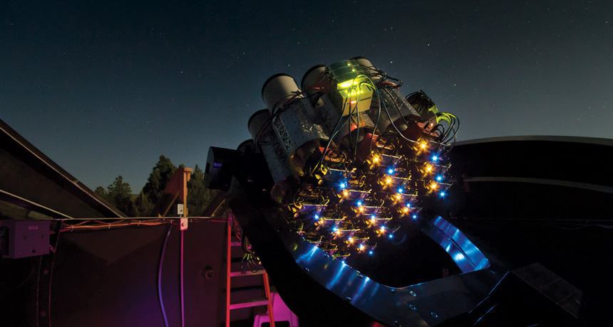
Astronomers can’t resist a good cosmic mystery. With detections of these galactic oddballs piling up, there is a push to figure out just how many of these things are out there and where they’re hiding. “There are more questions than answers,” says Remco van der Burg, an astrophysicist at CEA Saclay in France. Cracking the code of dark galaxies could provide insight into how all galaxies, including the Milky Way, form and evolve. Compound eye on the sky Telescopes designed to detect faint objects have revealed the presence of many sizable but near-empty galaxies — officially known as “ultradiffuse galaxies.” The deluge of discoveries started in New Mexico, with a telescope that looks more like a honeycomb than a traditional observatory. Sitting in a park about 110 kilometers southwest of Roswell (a city that has turned extraterrestrials into a tourism industry), the Dragonfly telescope consists of 48 telephoto lenses; it started with three in 2013 and continues to grow. The lenses are divided evenly among two steerable racks, and each lens is hooked up to its own camera. Partly inspired by the compound eye found in dragonflies and other insects, this relatively small scope has revealed dim galaxies missed by other observatories.
The general rule for telescopes is that bigger is better. A large mirror or lens can collect more light and therefore see fainter objects. But even the biggest telescopes have a limitation: unwanted light. Every surface in a telescope is an opportunity for light coming in from any direction to reflect onto the image. The scattered light shows up as dim blobs, or “ghosts,” that can wash out faint detail in pictures of space or even mimic very faint galaxies.
Large dark galaxies look a lot like these ghosts, and so went unnoticed. But Dragonfly was designed to keep these splashes of light in check. Unlike most conventional professional telescopes, it has no mirrors. Precision antireflection coatings on the lenses keep scattered light to a minimum. And having multiple cameras pointed at the same part of the sky helps distinguish blobs of light bouncing around in the telescope from blobs that actually sit in deep space. If the same blob shows up in every camera, it’s probably real.
“It’s a very clever idea, very brilliant,” says astronomer Jin Koda of Stony Brook University in New York. “Dragonfly made us realize that there is a chance to find a new population of galaxies beyond the boundary of what we know so far.”
In spring 2014, researchers pointed Dragonfly at the well-studied Coma cluster, a conglomeration of thousands of galaxies. At a distance of about 340 million light-years, Coma is a close, densely packed collection of galaxies and a rich hunting ground for astronomers. A team led by Abraham and astronomer Pieter van Dokkum of Yale University was looking at the edges of galaxies for far-flung stars and stellar streams, evidence of the carnage left behind after small galaxies collided to build larger ones. They were not expecting to find dozens of galaxies hiding in plain sight. “People have been studying Coma for 80 years,” Abraham says. “How could we find anything new there?” And yet, scattered throughout the cluster appeared 47 dark galaxies, many of them comparable in size to the Milky Way — tens of thousands to hundreds of thousands of light-years across (SN: 12/13/14, p. 9). This was perplexing. A galaxy that big should have no problem forming lots of stars, van Dokkum and colleagues noted in September in Astrophysical Journal Letters.
Hidden strength Even more surprising, says Abraham, is that those galaxies survive in Coma, a cluster crowded with galactic bullies. A galaxy’s own gravity holds it together, but gravity from neighboring galaxies can pull hard enough to tear apart a smaller one. To create sufficient gravity to survive, a galaxy needs mass in the form of stars, gas and other cosmic matter. In a place like Coma, a galaxy needs to be fairly massive or compact. But with so few stars (and presumably so little mass) spread over a relatively large space, dark galaxies should have been shredded long ago. They are either recent arrivals to Coma or a lot stronger than they appear.
From what researchers have learned so far, dark galaxies seem to have been lurking for many billions of years. They are located throughout their home clusters, suggesting that they’ve had a long time to spread out among the other galaxies. And the meager stars they have are mostly red, indicating that they are very old. With this kind of longterm survival, dark galaxies probably have a hidden strength, most likely due to dark matter.
All galaxies are loaded with dark matter, a mysterious substance that reveals itself only via gravitational interactions with luminous gas and stars. Much of that dark matter sits in an extended blob (known as the halo) that reaches well beyond the visible edge of a galaxy. On average, dark matter accounts for about 85 percent of all the matter in the universe. Within the central regions of the dark galaxies in Coma, dark matter must make up about 98 percent of the mass for there to be enough gravity to keep the galaxy intact, van Dokkum and colleagues say. Dark galaxies appear to have similar fractions of dark matter focused near their cores as the Milky Way does throughout its broader halo.
Astronomers had never seen such a strong preference for dark matter in galaxies so large. The initial cache of galactic enigmas lured a slew of researchers to the hunt. They pored over existing images of Coma and other clusters, looking for more dark galaxies. These galaxies are so faint that they could easily blend in with a cluster’s background light or be mistaken for reflections within a telescope. But once the galaxy hunters knew what to look for, they were not disappointed — those first 47 were just the tip of the iceberg.
Looking at old images of Coma taken by the Subaru telescope in Hawaii, Koda and colleagues easily confirmed that those 47 were really there. But that wasn’t all. They found a total of 854 dark galaxies, 332 of which appeared to be roughly the size of the Milky Way (SN: 7/25/15, p. 11). They calculated that Coma could harbor more than 1,000 dark galaxies of all sizes — comparable to its number of known galaxies. Astronomer Christopher Mihos of Case Western Reserve University in Cleveland and colleagues, reporting in 2015 in Astrophysical Journal Letters, found three more in the Virgo cluster, a more sparsely populated but closer gathering of galaxies that’s a mere 54 million light-years away.
In June, van der Burg and collaborators reported another windfall in Astronomy & Astrophysics. Using the Canada-France-Hawaii Telescope atop Mauna Kea in Hawaii, they measured the masses of several galaxy clusters. Taking a closer look at eight clusters, all less than about 1 billion light-years away, the group found roughly 800 more ultradiffuse galaxies.
“As we go to bigger telescopes, we find more and more,” says Michael Beasley, an astrophysicist at Instituto de Astrofísica de Canarias in Santa Cruz de Tenerife, Spain. “We don’t know how many there are, but we know there are a lot of them.” There could even be more dark galaxies than bright ones.
Nature vs. nurture What dark galaxies are and how they formed is still a mystery. There are many proposals, but with so little data, few conclusions. For the vast majority of dark galaxies, researchers know only how big and how bright each one is. Three so far have had their masses measured. Of those, two appear to have more in common masswise with some of the small galaxies that orbit the Milky Way, while the third is as massive as our galaxy itself — roughly 1 trillion times as massive as the sun.
A dark galaxy in the Virgo cluster, VCC 1287, and another in Coma, Dragonfly 17, each have a total mass of about 70 billion to 90 billion suns. But only about one one-thousandth of that or less is in stars. The rest is dark matter. That puts the total masses of these two galaxies on par with the Large Magellanic Cloud, the largest of the satellite galaxies that orbit the Milky Way. But focus on just the mass of the stars, and the Large Magellanic Cloud is about 35 times as large as Dragonfly 17 and roughly 100 times as large as VCC 1287.
A galaxy dubbed Dragonfly 44, however, is another story. It’s a dark beast, weighing about as much as the entire Milky Way and made almost entirely of dark matter, van Dokkum and colleagues report in September in Astrophysical Journal Letters. “It’s a bit of a puzzle,” Beasley says. “If you look at simulations of galaxy formation, you expect to have many more stars.” For some reason, this galaxy came up short. The environment may be to blame. A cluster like Coma grows over time by drawing in galaxies from the space around it. As galaxies fall into the cluster, they feel a headwind as they plow through the hot ionized gas that permeates the cluster. The headwind can strip gas from an incoming galaxy. But galaxies need gas to form stars, which are created when self-gravity crushes a blob of dust and gas until it turns into a thermonuclear furnace. If a galaxy falls into the cluster just as it is starting to make stars, this headwind might remove enough gas to prevent many stars from forming, leaving the galaxy sparsely populated.
Or maybe there’s something intrinsic to a galaxy that turns it dark. A volley of supernovas or a prolific burst of star formation might drive gas out of the galaxy. Nicola Amorisco of the Max Planck Institute for Astrophysics in Garching, Germany, and Abraham Loeb of the Harvard-Smithsonian Center for Astrophysics in Cambridge, Mass., suggest that ultradiffuse galaxies start off as small galaxies that spun rapidly as they formed. All galaxies rotate, but perhaps dark galaxies are a subset that twirl so fast that their stars and gas have spread out, turning them into diffuse blobs rather than star-building machines.
To test these and other ideas, astronomers are focused on two key pieces of information: the masses of these galaxies and their locations in the universe. Mass can help researchers distinguish between formation scenarios, such as whether or not dark galaxies are failed Milky Way–like behemoths. A survey of other locales would indicate whether dark galaxies are unique to big clusters such as Coma, suggesting that the environment plays a role in their creation. But if they turn up outside of clusters, isolated or with small groups of galaxies, then perhaps they’re just born that way.
There’s already a hint that dark galaxies depend more on nature than nurture. Yale astronomer Allison Merritt and colleagues reported in October online at arXiv.org that four ultradiffuse galaxies lurk in a small galactic gathering about 88 million light-years away, indicating that clusters aren’t the only place dark galaxies can be found. And van der Burg, in his survey of eight clusters, found that dark galaxies make up the same fraction of all galaxies in a cluster regardless of cluster mass — at least, for clusters weighing between 100 trillion and 1 quadrillion times the mass of the sun. About 0.2 percent of the mass of the stars is tied up in the dark galaxies. Since all eight clusters host roughly the same relative number of dark galaxies, that suggests that there is something intrinsic about a galaxy that makes it dark, van der Burg says.
What this all means for understanding how galaxies form is hard to say. These cosmic specters might be an entirely new entity that will require new ideas about galaxy formation. Or they could be one page from the galaxy recipe book. Timing, location and luck might send some of our heavenly neighbors toward a bright future and force others to fade into the background. Perhaps dark galaxies are a mixed bag, the end result of many different processes going on in a variety of environments.
“I see no reason why the universe couldn’t make these things in many ways,” Abraham says. “Part of the fun over the next few years will be to figure out which is in play in any particular galaxy and what sort of objects the universe has chosen to make.”
What is clear is that as astronomers push to new limits — fainter, farther, smaller — the universe turns up endless surprises. Even in Coma, a locale that has been intensively studied for decades, there are still things to discover. “There’s just a ton of stuff out there that we’re going to find,” Abraham says. “But what that is, I don’t know.”